
Microscopes enhance our capabilities to ‘see’ objects of micron size dimensions. The resolving capability of a microscope depends on the wavelength of the illumination. We have developed microscopes that use radiation with wavelengths in the range 13-47 nm for illumination. These short wavelengths have allowed these microscopes to image nanoscale structures on surfaces. The high peak power of the extreme ultraviolet laser output enables to capture images with a single shot. In combination, the microscopes can take sequential images that map dynamic phenomena.
46.9 nm Microscope
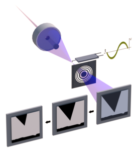
The microscope schematically shown in Fig. 1 uses the output of a compact 46.9 nm laser for illumination. This microscope was used to image carbon nanotubes with single laser shot at wavelength resolution (Fig. 2). Its capability for single shot imaging enabled the microscope to follow the repetitive motion of a cantilever oscillating at tens of kHz repetition rate (Fig. 2). This microscope can also operate in reflection configuration making it possible to image nanopatterns on surfaces.

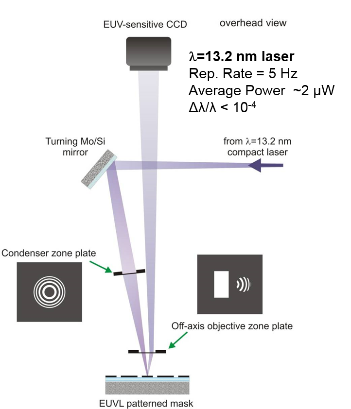
Aerial Mask Inspection at 13.2 nm

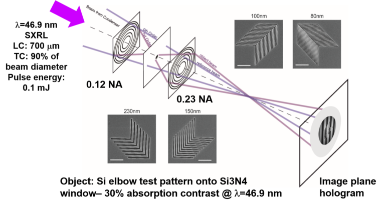
Holographic Imaging Microscopy
Selected Publications
- G. Vaschenko, F. Brizuela, C. Brewer, Y. Wang, M.A. Larotonda, B.M. Luther, M.C. Marconi, J.J. Rocca, C.S. Menoni, E.H. Anderson, W. Chao, B.D. Harteneck, J.A. Liddle, Y. Liu, and D.T. Attwood, “Sub-38 nm resolution tabletop microscopy with 13 nm wavelength laser light”, Optics Letters 31, 1214, (2006).
- Brizuela, S. Carbajo, A. Sakdinawat, D. Alessi, D. H. Martz, Y. Wang, B. Luther, K. A. Goldberg, I. Mochi, D. T. Attwood, B. La Fontaine, J. J. Rocca, and C. S. Menoni, “Extreme ultraviolet laser-based table-top aerial image metrology of lithographic masks,” Opt. Express 18, 14467-14473 (2010).
- S. Carbajo, I. Howlett, F. Brizuela, K. S. Buchanan, M. C. Marconi, W. Chao, E. H. Anderson, I. Artioukov, A. Vinogradov, J.J. Rocca, and C.S. Menoni, “Sequential single-shot imaging of nanoscale dynamic interactions with a table-top soft x-ray laser,“ Opt. Letters, 37, pp. 2994-2996 (2012).
- Nejdl; ID Howlett; D. Carlton; E.H. Anderson; W. Chao; M.C. Marconi; J.J. Rocca; C.S. Menoni, “Image Plane Holographic Microscopy With a Table-Top Soft X-Ray Laser,” IEEE Photonics Journal 7 (1); Article Number: 6900108, (2015). DOI: 10.1109/JPHOT.2015.2389957.
Funding
National Science Foundation, Extreme Ultraviolet Science and Technology Center